Health
New Holographic Optogenetics Techniques Transform Brain Mapping
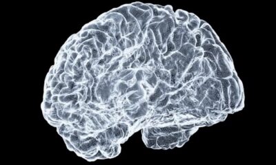
Recent advances in neuroscience research are set to revolutionize the way scientists map the brain’s structure and synaptic connections. Two distinct research teams have developed innovative techniques that combine holographic optogenetics with advanced computational methods, significantly enhancing the speed and accuracy of mapping synapses in living brains.
One research team, comprised of scientists from Columbia University and UC Berkeley, published their findings in the journal Nature Neuroscience. Led by Marcus A. Triplett, the group aimed to create tools that address the pressing need for high-resolution mapping of neuronal connections. “Understanding how the nervous system is wired is important because that wiring is a large part of what gives the brain’s circuitry its function,” Triplett explained.
Despite the advancements in imaging techniques, traditional methods like electron microscopy have limitations. While they produce detailed images of fixed brain tissue, they cannot provide real-time insights into the dynamic connections between neurons in living organisms. Triplett emphasized the necessity for a technique that could not only map large volumes of brain tissue but also measure crucial variables, such as the strength of neuronal connections.
To achieve this, the researchers employed holographic optogenetics, a method that uses light to selectively activate specific neurons. By introducing light-sensitive proteins called opsins into the neurons, they demonstrated a method of recording electrical activity between neurons. This innovative approach allowed them to determine if a synaptic connection existed based on the transmission of neural activity between the activated and recorded neurons.
The results were remarkable. The new technique enabled Triplett’s team to map ten times more connections than previously possible within the same timeframe. “Our technique will see the greatest use in studying neural computation, revealing how the brain’s wiring supports its computational abilities,” Triplett noted.
At the same time, the “Wavefront Engineering Microscopy team” at Sorbonne University has been exploring similar applications of holographic optogenetics for real-time neuronal mapping. Led by Dimitrii Tanese, the researchers focused on creating optical tools that allow precise control of neuronal activity without invasive methods. Tanese remarked, “We sought to establish and validate a scalable framework for mapping synaptic connections directly in the intact brain with high precision and speed.”
Conventional approaches to mapping neuronal connections often involve invasive electrode implantation, which limits the ability to observe multiple connections simultaneously. Tanese’s team utilized two-photon holographic stimulation, which allows for precise targeting of specific cells using light. This method can activate multiple neurons at once, effectively performing group testing, which accelerates the mapping process.
Their findings indicate that the new approach can map connections among up to 100 presynaptic neurons in a live mouse brain within just five minutes—an impressive improvement over previous methods. “By combining speed, precision, and scalability, our approach overcomes the main limitations of traditional techniques,” Tanese stated.
The implications of these advancements extend beyond basic research. Understanding how individual neurons connect within a living brain could elucidate crucial aspects of brain function, including how the brain reorganizes during learning and recovery from injury. Both teams are now working to refine their techniques further, with Tanese’s group exploring the integration of voltage indicators to enhance sensitivity in detecting neuronal signals.
As they continue their research, Triplett and his colleagues are particularly excited about the potential applications in studying visual perception and are scaling their techniques to map larger populations of neurons. “One cubic millimeter of the mouse brain contains tens to hundreds of thousands of neurons. There’s a lot of work to do!” Triplett remarked.
In summary, the development of these holographic optogenetics techniques marks a significant leap forward in neuroscience, paving the way for more comprehensive understanding of brain connectivity and function. As researchers continue to refine these methods, the potential for breakthroughs in understanding neurological disorders and brain functionality grows ever more promising.

Homeowner Faces Foreclosure Over $121K ‘Zombie Debt’ Resurgence

Urgent Update: SNAP Benefits Cut to 50% for Millions in November

Burger King’s 2025 Advent Calendar Launches Soon After Last Year’s Sellout

Azerbaijan Faces Increased Scrutiny Over Sanctions Compliance

Innovative AI System Monitors Driver Stress Levels for Safety

Renowned Sportscaster Michael Barkann to Speak at Rodeph Shalom

Nvidia and Tech Stocks Boost Wall Street Amid Broader Decline

Researchers Develop Sustainable Sodium-Ion Batteries from Lignin

Philadelphia’s Michael Barkann to Share Sports Insights at Rodeph Shalom

Researchers Challenge 200-Year-Old Physics Principle with Atomic Engines

Hamas Chief Stresses Disarmament Tied to Occupation’s End

Syracuse Stage Delivers Lively Adaptation of ‘The 39 Steps’

Federal Agents Detain Driver in Addison; Protests Erupt Immediately

Ohio State Study Uncovers Brain Connectivity and Function Links

Global Military Spending: Air Forces Ranked by Budget and Capability

NHP Foundation Secures Land for 158 Affordable Apartments in Denver

Trump’s Push to Censor National Parks Faces Growing Backlash

NFL Confirms Star-Studded Halftime Show for Super Bowl LVIII
-
 Science3 weeks ago
Researchers Challenge 200-Year-Old Physics Principle with Atomic Engines
-
 Politics1 week ago
Hamas Chief Stresses Disarmament Tied to Occupation’s End
-
 Entertainment1 week ago
Syracuse Stage Delivers Lively Adaptation of ‘The 39 Steps’
-
 Top Stories1 week ago
Federal Agents Detain Driver in Addison; Protests Erupt Immediately
-
 Science7 days ago
Ohio State Study Uncovers Brain Connectivity and Function Links
-
 World3 weeks ago
Global Military Spending: Air Forces Ranked by Budget and Capability
-
 Politics2 weeks ago
NHP Foundation Secures Land for 158 Affordable Apartments in Denver
-
 Lifestyle1 week ago
Trump’s Push to Censor National Parks Faces Growing Backlash
-
 Politics1 week ago
NFL Confirms Star-Studded Halftime Show for Super Bowl LVIII
-

 World1 week ago
Boeing’s Aircraft Production: Assessing Numbers and Challenges
-

 Top Stories1 week ago
Will Smith Powers Dodgers to World Series Tie with Key Homer
-

 Lifestyle1 week ago
Red Bluff High School’s Elli Nolan Named Rotary Student of the Month