Science
Scientists Map Fetal Heart Development, Uncover Congenital Defect Insights
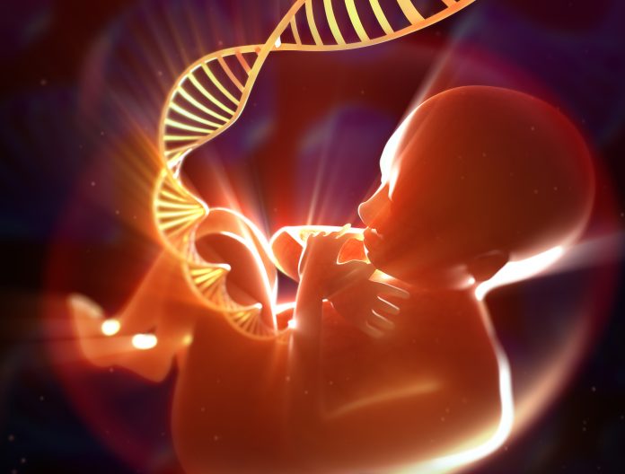
Researchers from the KTH Royal Institute of Technology have developed what they describe as the “most comprehensive spatiotemporal atlas of first-trimester human heart development to date.” Their findings, published in the journal Nature Genetics, provide crucial insights into how various cells interact and organize during the critical phases of fetal heart development. This work could significantly enhance prenatal care and lead to innovative treatments for congenital heart defects.
The study, titled “Spatiotemporal gene expression and cellular dynamics of the developing human heart,” presents a detailed blueprint of the heart’s development during the late first and early second trimesters. It highlights how essential structures such as the pacemaker system, heart valves, and the wall between the heart’s upper chambers form and function. According to Enikő Lázár, MD, PhD, a postdoctoral researcher in the Department of Gene Technology at KTH Royal Institute of Technology, “Understanding how the cardiac architecture forms could improve prenatal care as well as lead to new treatments for congenital defects.”
Key Discoveries on Cardiac Development
Among the significant findings is the identification of a previously unknown group of cells that produce adrenaline, termed “neuroendocrine chromaffin” cells. Lázár notes that these cells may be unique to humans and could aid the heart’s response to low oxygen levels during development or birth. Their role in coordinating the fetal heart’s response to stress is critical, and they may also indicate a cellular origin for cardiac pheochromocytomas, rare tumors that develop in the heart.
The research further uncovers the diversity of cells constituting the heart valves and atrial septum, providing insights into the formation of the heart’s internal structures and the reasons behind congenital defects such as valve malformations. The study also highlights the presence of diverse supporting cells, or mesenchymal cells, that contribute to the heart’s structural integrity and may be involved in conditions like valve defects or arrhythmias.
Another pivotal aspect of the research is the mapping of the intricate wiring of cells that form the heart’s natural pacemaker and conduction system. This includes the sinoatrial node, which regulates heartbeat, and the Purkinje fibers, which distribute electrical signals throughout the heart. The study reveals how nerve and support cells connect with pacemaker cells and indicates that different types of neurotransmitters begin to influence heart development early on.
Future Implications and Interactive Tools
While the findings are groundbreaking, the researchers acknowledge limitations in their study, noting that it does not encompass the first two weeks of cardiogenesis. During this period, many genes related to congenital heart disease become active. They emphasize that increasing the sample size could enhance the resolution of their analysis and enable further validation of the identified cell populations.
The team has made their findings accessible through an interactive online tool, allowing scientists to explore deeper insights into early cardiogenesis. The researchers believe that their datasets can provide valuable references for early expression patterns of candidate genes linked to congenital heart diseases and for benchmarking models derived from human pluripotent stem cells.
In conclusion, the work by the KTH Royal Institute of Technology not only sheds light on the complexities of fetal heart development but also offers hope for improved understanding and treatment of congenital heart defects.

Baxter International Faces Class Action as Investors Seek Lead Role

Netflix’s ‘The Perfect Neighbor’ Redefines True Crime Genre

Psychotherapist Offers Key Strategies for Parents to Revive Intimacy

San Diego Food Banks Mobilize as CalFresh Benefits Face Delays

New Mexico Launches Free Universal Child Care Program Next Week

Texas Voters Face Crucial Decision on $3 Billion Dementia Research Fund

Immigration Enforcement Drives Chicago Health Care Crisis

FAA Issues Urgent Ground Stop at Newark Airport Amid Shutdown

CU Buffs Urgently Address Run Defense After 422-Yard Loss

Researchers Challenge 200-Year-Old Physics Principle with Atomic Engines

NHP Foundation Secures Land for 158 Affordable Apartments in Denver

Boeing’s Aircraft Production: Assessing Numbers and Challenges

Syracuse Stage Delivers Lively Adaptation of ‘The 39 Steps’

Neuroscientist Advocates for Flag Football Until Age 14

Longtime Friends Face Heartbreak After Loss and Isolation

Trump’s Push to Censor National Parks Faces Growing Backlash

Global Military Spending: Air Forces Ranked by Budget and Capability

Red Bluff High School’s Elli Nolan Named Rotary Student of the Month
-
 Science2 weeks ago
Researchers Challenge 200-Year-Old Physics Principle with Atomic Engines
-
 Politics2 weeks ago
NHP Foundation Secures Land for 158 Affordable Apartments in Denver
-
 World5 days ago
Boeing’s Aircraft Production: Assessing Numbers and Challenges
-
 Entertainment4 days ago
Syracuse Stage Delivers Lively Adaptation of ‘The 39 Steps’
-
 Health2 weeks ago
Neuroscientist Advocates for Flag Football Until Age 14
-
 Lifestyle2 weeks ago
Longtime Friends Face Heartbreak After Loss and Isolation
-
 Lifestyle4 days ago
Trump’s Push to Censor National Parks Faces Growing Backlash
-
 World2 weeks ago
Global Military Spending: Air Forces Ranked by Budget and Capability
-
 Lifestyle5 days ago
Red Bluff High School’s Elli Nolan Named Rotary Student of the Month
-

 Science4 days ago
AI Misidentifies Doritos Bag as Gun, Triggers Police Response
-

 Business2 weeks ago
Spirit Airlines Cuts Workforce with Furloughs for 365 Pilots
-

 Top Stories5 days ago
Urgent Search for Suspect Who Exposed Himself to Teen Girl